Surgical technology & navigation
Ultrasonic bone scalpel
Misonix BoneScalpel
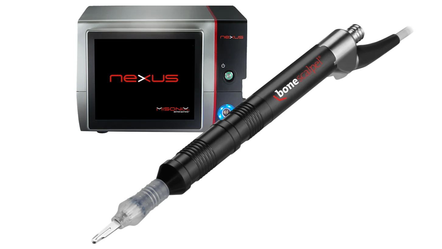
The bone scalpel is an ultrasonic blade that vibrates at high frequency over a very short distance. It cuts mineralized bone efficiently but is much gentler on soft tissue than a high-speed burr, so it can be used closer to the dura and nerve roots during a laminectomy, osteotomy or decompression.
Spinal navigation
CT-guided navigation
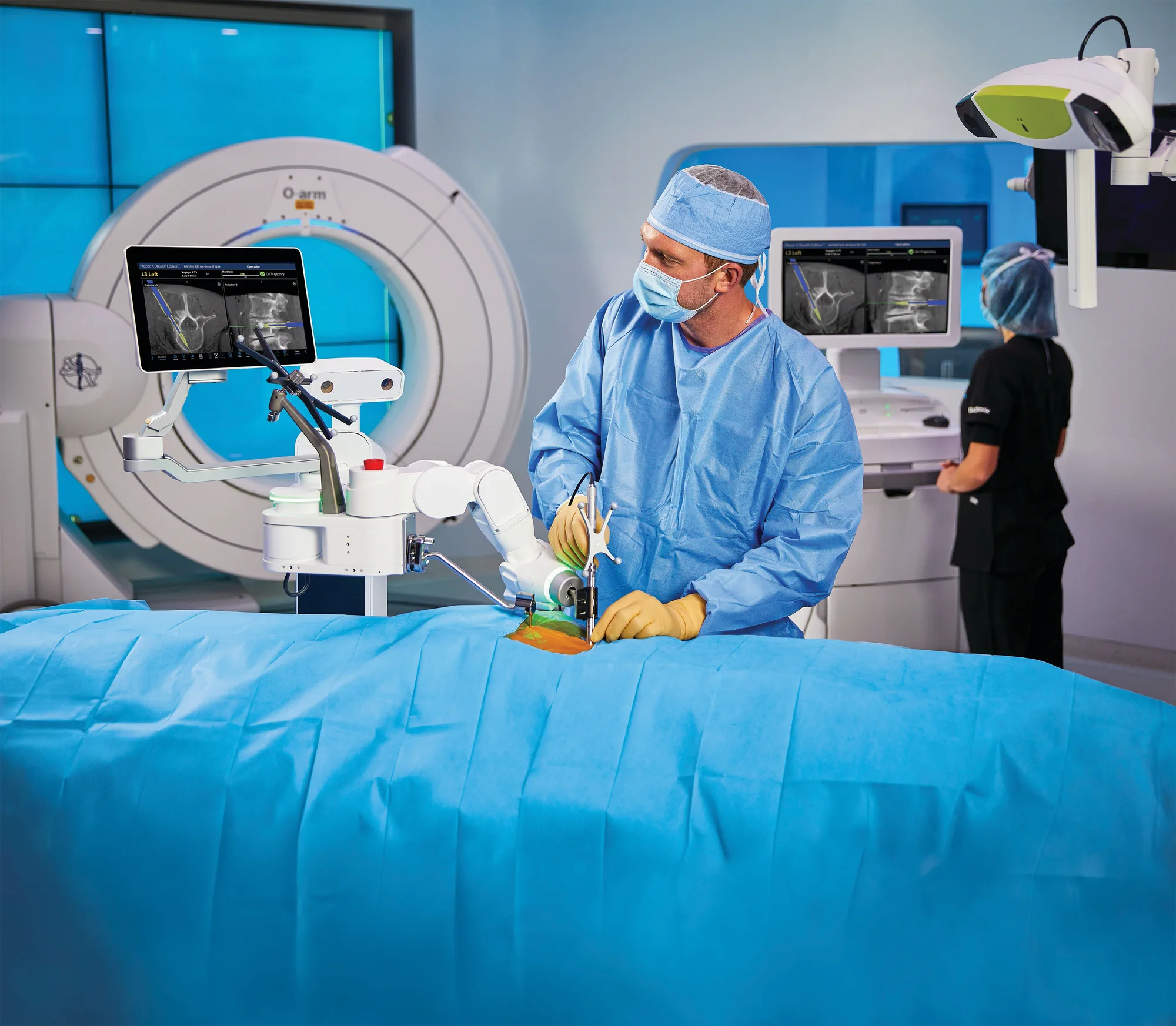
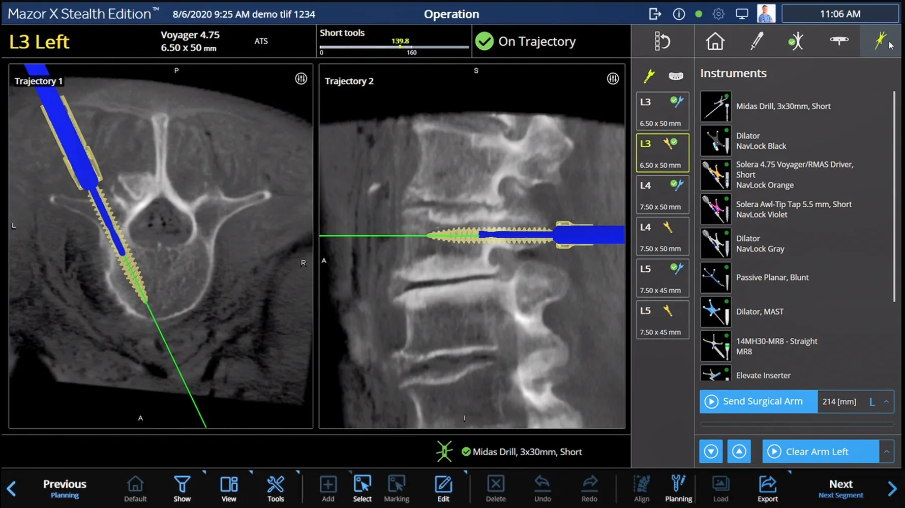
Instruments are tracked live against a CT scan taken in the operating room once you are positioned for surgery, so the path of each screw is checked against your anatomy as it lies on the table.
Intraoperative spinal robot
Globus ExcelsiusGPS and Medtronic Mazor
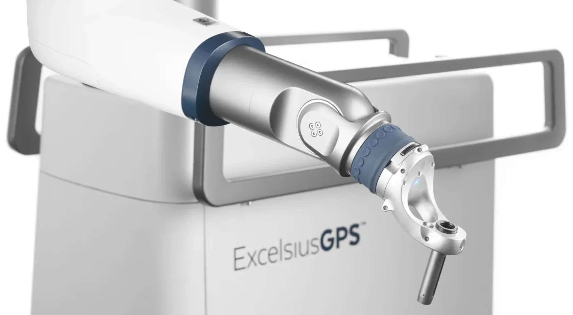
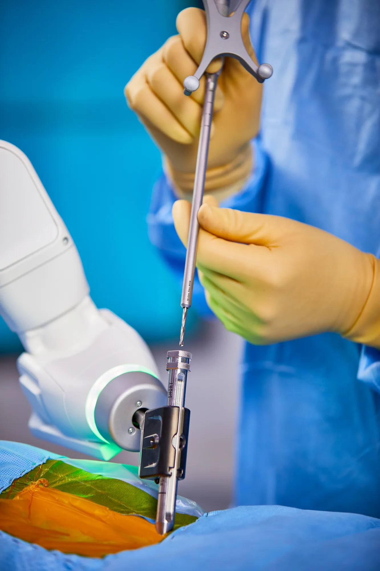
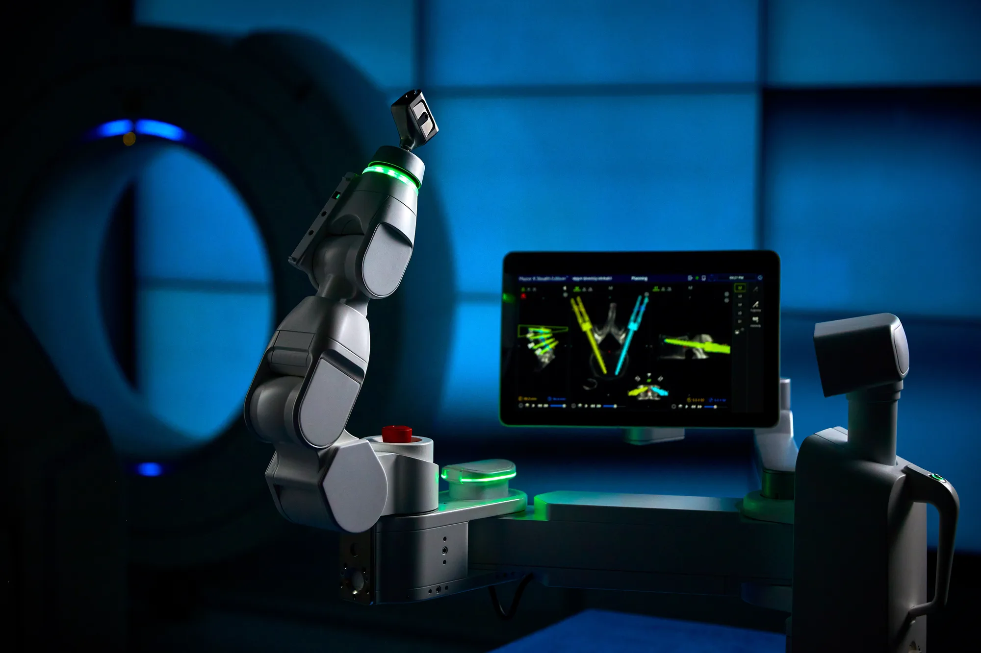
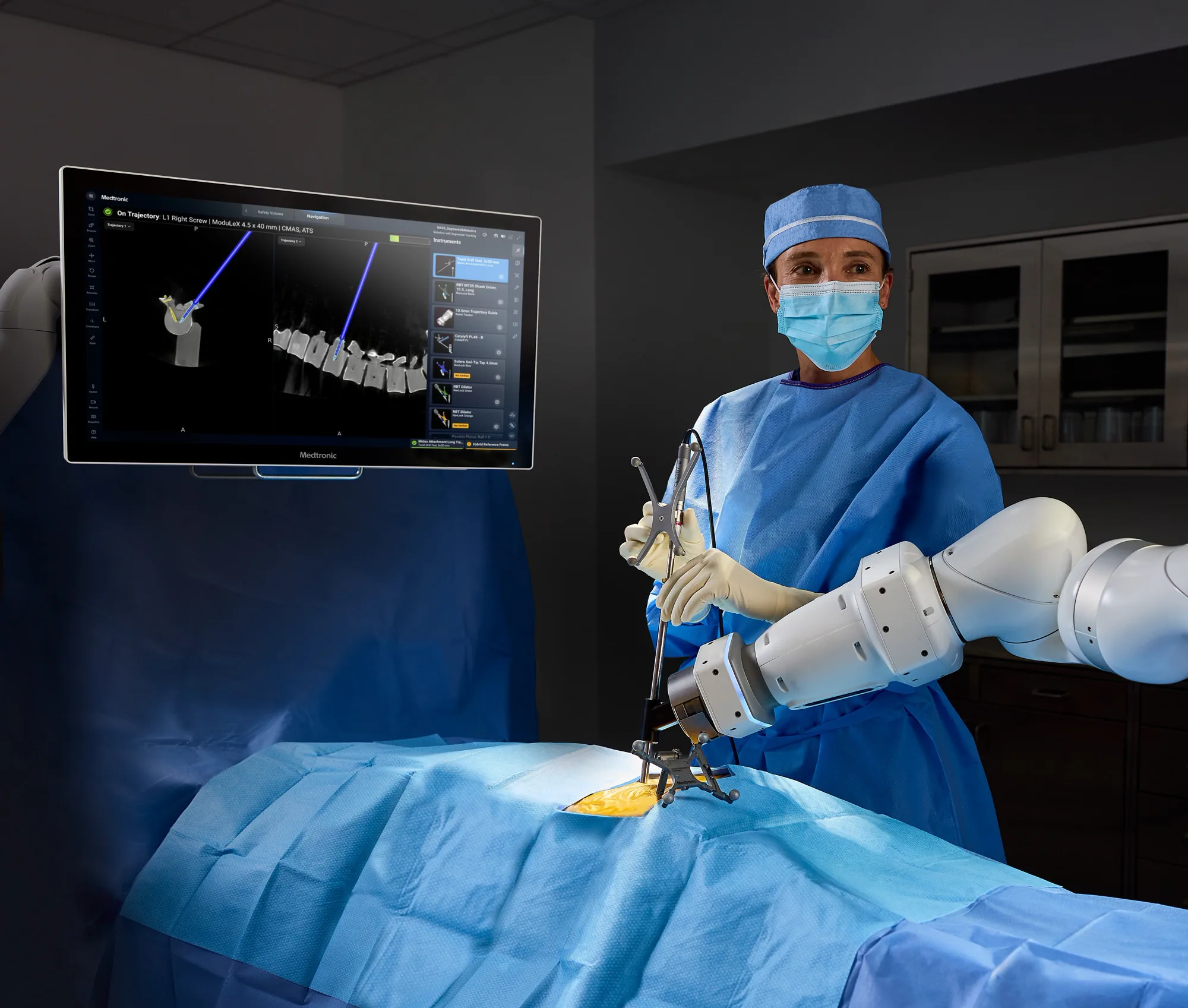
A robotic arm holds the planned path steady while the screw is placed. This stops the instrument drifting, as it can when guided by hand along dense bone, and helps most in minimally invasive surgery, where the anatomy cannot be seen directly. Several companies make spinal robots, including Globus and Medtronic, and they all work this way.
Intraoperative CT scan
Medtronic O-arm
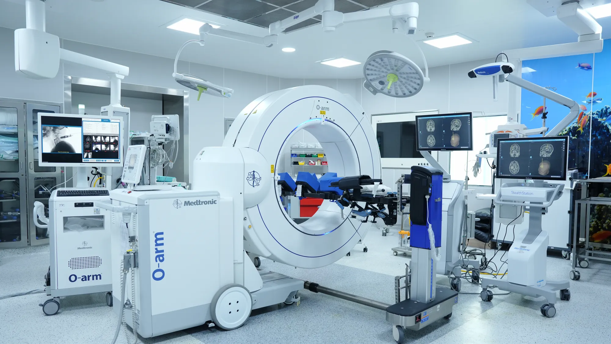
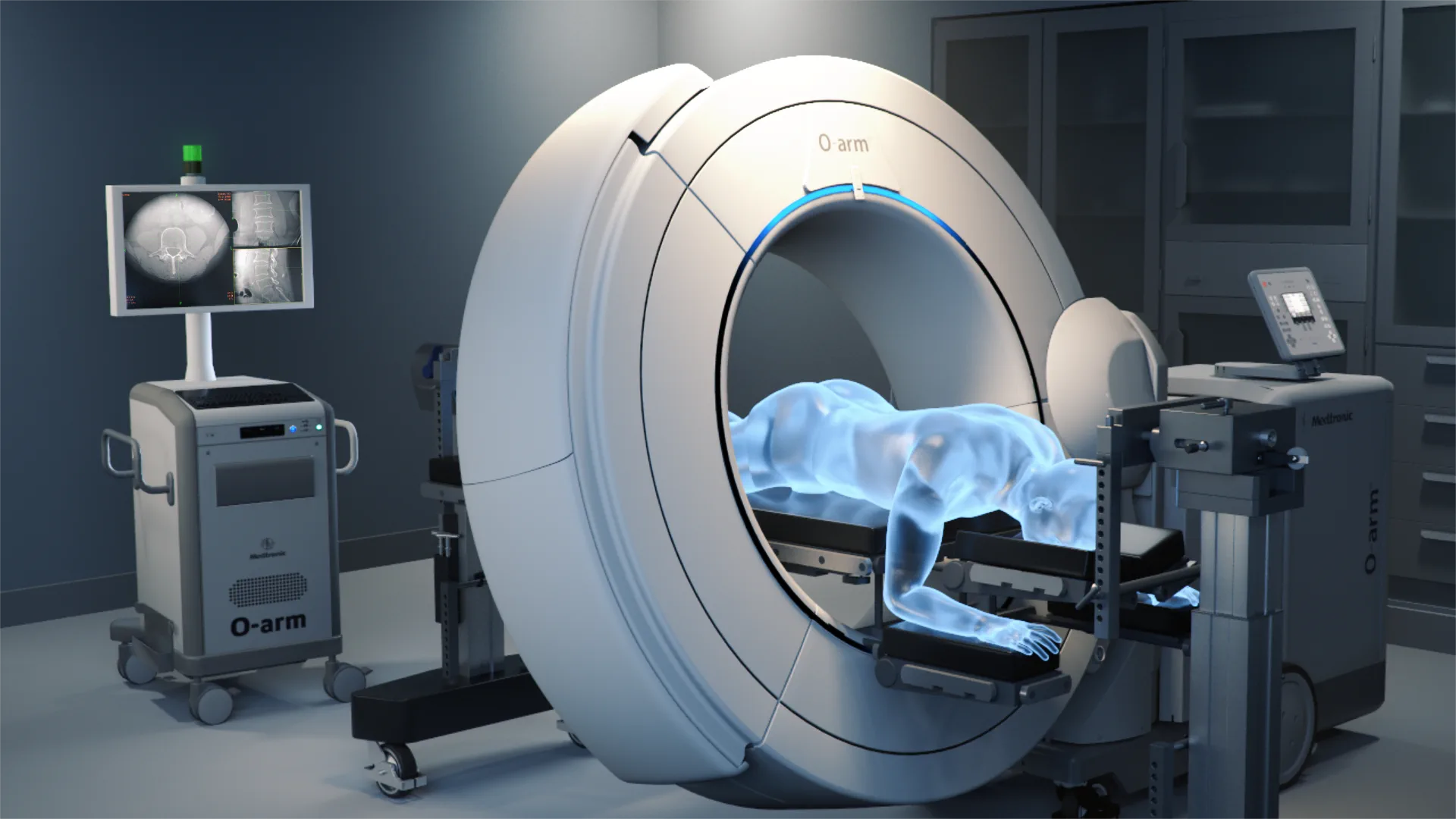
The O-arm takes a 3D CT scan in the operating room once you are positioned for surgery. The navigation then works from your anatomy exactly as it lies on the table.
Neuromonitoring
EMG, motor and sensory evoked potentials
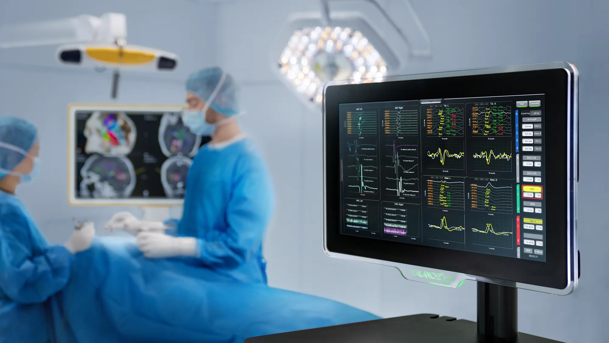
- SSEP
- Monitors sensation from the nerve to the brain. Signal change threshold: 50% smaller amplitude or 10% longer latency.
- MEP
- Monitors motor pathways from the brain to the nerve. Signal change threshold: 50–80% smaller amplitude, or lost.
- EMG
- Monitors spontaneous activity within the nerve. Signal change threshold: sustained bursts of activity.
Neuromonitoring tracks the nerve pathways in real time. It can alert your surgeon when a nerve is being compressed, so corrective action can be taken, reducing the risk of permanent nerve injury.
A neurophysiologist monitors nerve and spinal cord function throughout the operation, so a change is caught while it is still reversible. Three signals are used together: EMG, from electrodes in the muscles each nerve root supplies, which can also test a screw directly for sitting too close to the nerve; motor evoked potentials, which watch the motor pathways of the spinal cord and matter most during cervical work near the cord; and somatosensory evoked potentials, which track the sensory pathways up the back of the cord. Monitoring lowers the chance that a nerve injury goes unnoticed, but it cannot remove the risk of one. Whether it is used depends on the procedure, the level and your anatomy.
Endoscopic spine surgery
Transforaminal endoscopic discectomy
The endoscope is a tube under a centimetre across that carries its own camera, light and saline, with a channel beside the camera for instruments. Passed from the side of the back into the opening where the nerve leaves the spine, it reaches the disc fragment without stripping muscle from the bone, and the fragment is removed under a magnified view on the screen. It suits a fragment that can be reached through that opening; not every herniation can be, and at L5–S1 the pelvis can block the approach from the side.